
From Businesswire:
BICMD Expands to Include All Major Medical Specialties
NEW YORK–(BUSINESS WIRE)–Best In Class MD (BICMD) announced its expansion of expert medical opinion (EMO) services to include 72 subspecialties within Internal Medicine, Surgery, Psychiatry, Radiology, Oncology and Obstetrics & Gynecology. This major initiative will double the company’s Expert Network by the end of 2022 to include over 350 fellowship-trained medical doctors.
The strategic decision to expand beyond orthopedics was made to meet demand of clients as well as the EMO market broadly. “We recognized that becoming a comprehensive offering would require representation across specialties, and we set out to recruit the highest quality and caliber of MDs to match the elite reputation we have built in the musculoskeletal space,” said Benedict Nwachukwu, MD, MBA, and Riley Williams, MD, BICMD’s co-founders and co-CEOs. “While musculoskeletal will remain a focus due to significant cost and disability, we appreciate the impact we can have as a multi-specialty organization that is more than a point solution.”
To showcase the breadth and quality of the new providers, BICMD is hosting a webinar on Wednesday, June 29, 4PM-5PM EST focused on an increasingly important area of need for their clients: Long Covid and its effects in the workforce. The increasing incidence of Long Covid has been defined as a “pandemic within a pandemic.” Although highly prevalent, there is limited understanding of Long Covid, resulting in limited access to experts who can guide management for payers and employers.
BICMD will demystify Long Covid with an expert panel moderated by Dr. Nwachukwu. Panelists include: Dr. Lawrence Purpura, Attending Infectious Disease Physician at NYP Columbia, Dr. Christina Eckhardt, Attending Pulmonology Physician at NYP Columbia, & Lara Heal, Senior workers compensation (WC) Case Manager at Maine Employers Mutual Insurance Company (MEMIC). They will discuss the pathophysiology of Long Covid, common questions WC claims teams are facing, and how Long Covid has impacted claims data. Attendees will have the opportunity to submit questions for a 30-minute Q&A segment. Registration is free, open to all, and a recording will be shared with those who register at the signup link.
Best In Class MD (BICMD) aims to improve the quality and appropriateness of care by ensuring that expert medical advice is at the center of care delivery. BICMD’s innovative telehealth platform creates a digital front door providing direct access to the nation’s elite medical minds.
Contacts:
Kirsten Grueter
Clinical Solutions
[email protected]
Orthopedic Shoulder, Elbow and Knee Specialist, Dr. Riley J. Williams III was once again awarded the Castle Connolly Top Doctors Award for 2022!
Dr. Riley J. Williams III is a member of an exclusive group of doctors that have met the physician-led Castle Connolly Top Doctor review process standards for at least 15 years.
Castle Connolly is the nations trusted source, for over 25 years in selecting Top Doctors across the country. Their intensive and lengthy vetting process includes peer nominations and Castle Connolly research. This physician-led team of researchers survey thousands of physicians and other healthcare professionals, asking them to identify doctors who are excellent in every specialty within their region throughout America.
Physicians cannot pay to become a Top Doctor and the award is only given to a very small percentage of the 850,000 physicians it considers. The Journal of Medical Research conducted a thorough study of patients, concluding that Castle Connolly’s peer-reviewed directory was more reliable than sites that relied only on patient reviews when it came to quality patient care. Dr. Riley J. Williams is honored to have received this prestigious award every year for the past fifteen years.
Congratulations to Dr. Riley J. Williams III, for once again, receiving the Castle Connolly’s 2022 Top Doctor Award!
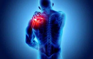
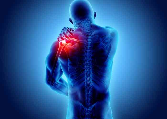
Shoulder Instability Doctor

Multidirectional shoulder instability can be caused by damage to the soft-tissue restraints within the shoulder. This type of damage can occur from repeated shoulder dislocations or from sports that require repetitive overhead motions, such as swimming, volleyball, tennis and baseball. Multidirectional shoulder instability can be surgically corrected by a shoulder capsulorrhaphy, if non-operative measures have failed to restore shoulder stability. Shoulder stability surgeon, Doctor Riley J. Williams provides diagnosis as well as surgical and nonsurgical treatment options for patients in Manhattan, Brooklyn, New York City and surrounding areas who are having issues with multidirectional shoulder instability. Contact Dr. Williams’ team today!
What is shoulder instability?
Shoulder instability describes the clinical scenario wherein the humeral head (upper arm bone) is able to slide out of the glenoid socket of the scapula (shoulder blade). Shoulder instability can result in a dislocating shoulder or a subluxating shoulder. Shoulder instability is typically described as anterior (front), inferior (bottom) or posterior (rear); the classification of shoulder instability corresponds to the direction of the dislocating humeral head. Shoulder instability can occur following a traumatic dislocation, participation in repetitive throwing sports (i.e. pitching) or in association with a developmental predisposition to having loose ligament surrounding the joints.

What is multidirectional shoulder instability?
The glenohumeral joint of the shoulder, one of the more complex joints within the body, is formed into a ball-and-socket type joint where the head of the humerus (upper arm bone) meets the glenoid cavity of the scapula (shoulder blade). This ball-and-socket joint arrangement enables joint movement in multiple directions. In some cases, the ligaments, tendons, and labrum (fibrous cartilage) of the shoulder do not provide adequate stabilization of the humeral head relative to the glenoid socket. This loss of soft-tissue restraint can cause the humerus and scapula to partially or completely separate or slide apart. When these soft tissue restraints lose their competency (i.e., stretch out or tear), recurrent joint dislocations can occur. When this instability occurs in two or more directions, the patient is described as having multidirectional shoulder instability. Sports that require repetitive overhead movement (swimming, volleyball, tennis, baseball) can cause athletes to experience multidirectional shoulder instability because of the cumulative effect or repetitive micro-trauma associated with overhead arm motion.
What is the treatment for multidirectional shoulder instability?
Individuals with multidirectional shoulder instability are typically treated non-operatively. A combination of rest, ice, and non-steroidal anti-inflammatory medications (NSAIDs) can be used for pain management following an acute dislocation. Physical rehabilitation and shoulder girdle strengthening is the mainstay of treating MDI patients. The muscles of the shoulder girdle are dynamic stabilizers of the shoulder joint. When the shoulder joint muscles are strong, they provide support to the ligaments, tendons, and labrum. Strong rotator cuff and peri-scapular muscles are necessary to hold the shoulder joint in place during high load sporting activities.
Patients with recurrent shoulder instability, and those who fail nonoperative management of their shoulder instability condition are treated surgically. The goal of surgery is to reestablish the competency of the static stabilizers of the gleno-humeral joint. This is achieved by either reattaching a torn labral complex, tightening a loose joint capsule or both. Tensioning a loose shoulder caspule or ligament is also known as shoulder capsulorraphy. Shoulder capsulorraphy can be performed using arthroscopy or open surgery. Understanding the clinical circumstances underlying a patient’s unstable shoulder is critical to determining which approach is best to achieve a successful outcome following surgery. Dr. Riley J. Williams, orthopedic shoulder doctor, treats patients in Manhattan, Brooklyn, New York City, NY and surrounding areas, who have experienced multidirectional shoulder instability and need a surgical repair.
How is a shoulder capsulorrhaphy performed?
Dr. Williams performs both arthroscopic and open shoulder capsulorraphy. Most individuals can be treated using minimally invasive techniques. This minimally invasive procedure involves small incisions to introduce a small camera (arthroscope) for Dr. Williams to methodically examine the muscles, tendons, ligaments, and cartilage of the shoulder joint. Specialized surgical instruments are inserted through another small incision and used to excise and remove the damaged tissues. The remaining healthy tendons, ligaments, and cartilage are wrapped around the shoulder joint to form a capsule. This capsule is subsequently fastened to the glenoid cavity of the scapula with special surgical anchors that are secured within the bone. These surgical anchors tighten the new shoulder capsule and realign the joint back to its correct anatomical position.
Those athletes who participate in contact sports (american football, rugby, lacrosse, wrestling) may be best treated using open shoulder capsulorrhaphy. The rate of re-dislocation in this group of individuals is lower following open surgery compared to arthroscopic repair techniques. The open surgical approach, known as open shoulder stabilization, requires a slightly larger incision to facilitate surgical access to the unstable ligament of the shoulder joint. Dr. Williams may recommend open surgery over an arthroscopic procedure for patients who have experienced bone loss, chronic multidirectional shoulder instability, or a failed shoulder joint reduction.
What is the recovery period like after a shoulder capsulorrhaphy?
The recovery period after a shoulder capsulorrhaphy is as follows:
- Joint immobilization using a sling for approximately 2 weeks following surgery.
- Pain and inflammation are managed with sling wear, ice, non-steroidal anti-inflammatory medications (NSAIDs), and for a brief period, narcotic pain medications.
- Physical therapy program will focus on strengthening the shoulder joint muscles and starts one week following surgery. Patient should expect to do PT for 2-3 months following the procedure.
- A full recovery is anticipated by 3-4 months. Contact sports are allowed 5+ months following surgery.
For more information on shoulder capsulorrhaphy, or to discuss your multidirectional shoulder instability treatment options, please contact the office of Riley J. Williams, MD, orthopedic shoulder doctor at the Hospital for Special Surgery (HSS), serving Manhattan, Brooklyn, New York City, NY and surrounding areas.

Biologic Treatments
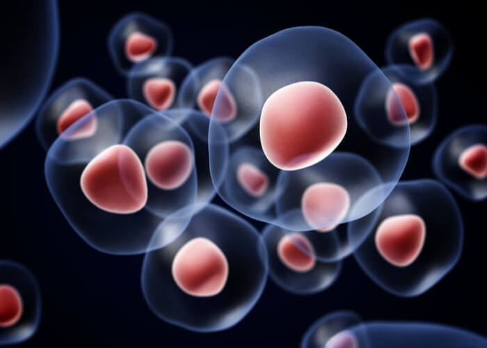
What are stem cells?
Stem cells naturally occur in the human body and possess the unusual ability to transform into any of a number of more specialized cells. There are two distinct properties that a cell must exhibit to be classified as a stem cell: self-renewal and potency. A cell’s ability to of remain undifferentiated after undergoing cellular division and cellular growth is known as self-renewal. Potency describes the cell’s ability to develop into more specialized cell. The study of these cellular properties has been seminal in advancing regenerative medicine techniques and methodologies, Dr. Riley J. Williams, orthopedic bone and joint preservation doctor, treats patients in Manhattan, Brooklyn, New York City, NY and surrounding areas, who are experiencing a bone or joint condition that can be treated with stem cell therapy.
Where are stem cells found?
In adults, stem cells can be found in adipose tissue, marrow blood, and peripheral blood.
What is stromal vascular fraction?
Stromal vascular fraction (SVF) is derived from adipose (fatty) tissue, contains a form of stem cell and is used for regenerative therapy. This material is found in lipoaspirate, a liquid by-product of liposuction, and contains a substantial number of adipose-derived stem cells. The stromal fraction of adipose tissue is the non-fatty component of what we know as human fat. Similar to stem cells harvested from bone marrow, adipose-derived stem cells are capable of multilineage differentiation. There are multiple cell types within the stromal vascular fraction including mesenchymal stem cells, B-cells, T-cells, lymphatic cells, and pericytes. Some orthopedic conditions can utilize these stem cells as an alternative form of regenerative therapy, including but not limited to:
- Arthritic conditions
- Ligament, tendon, and soft tissue injuries
- Cartilage injuries
- Meniscal tears
- Joint inflammation
- Muscle tears (strains)
How are adipose based stem cells harvested
The procedure to harvest these stem cells take approximately 20-30 minutes. A twilight anesthetic is administered to the patient prior to the procedure. Dr. Williams uses a needle to removed fat from the belly and flank area of the patient. Once the desired amount of whole fat has been removed, the stromal vascular fraction is separated from the lipoaspirate in a closed sterile system. The harvested adipose tissue undergoes multiple filtration protocols until the vascular fraction is isolated. The sterile, filtered stromal fraction is then injected into the injured area.
What are pericyte cells?
Pericyte cells can be found embedded in the membrane that surrounds capillaries and venules. The location of these cells allows them to play a significant role in the repair process by communicating with endothelial cells through paracrine signaling or direct contact. This cellular communication is the driving force behind vascular repair, the inflammatory response, constructing new blood vessels (angiogenesis), and the removal of toxins from the tissues.
These cells have long been known to play a significant role in the endothelial tissue repair process, but recent studies have shown pericyte cells to possess mesenchymal repair properties as well. Researchers have harnessed the potency and self-renewal characteristics of pericytes and developed a new method for stem cell therapy. This newer version of regenerative therapy has shown some promise in treating the following orthopedic conditions:
- Muscle injuries
- Cartilage injuries
- Ligament and tendon injuries
- Fractures
Similar to stromal vascular fraction, pericytes are harvested using lipoaspiration (liposuction). These cells are isolated using a closed filtration process. The resulting tissue graft is typically injected into the injured area of interest.
For more information on stem cell therapy, stromal vascular fraction, or to discuss your joint preservation treatment options, please contact the office of Riley J. Williams, MD, orthopedic bone and joint preservation doctor at the Hospital for Special Surgery (HSS), serving Manhattan, Brooklyn, New York City, NY and surrounding areas.
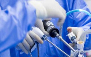
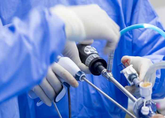
Elbow Arthroscopy Doctor
Do you have osteoarthritis or degenerative joint disease? Are you unable to bend or straighten your elbow due to stiffness or catching? If so, you may have bone spurs or osteophytes in your elbow. Elbow bone spurs, also called osteophytes can be caused by osteoarthritis, overuse, or trauma. Bone spurs can be removed non-invasively, with arthroscopic surgery. Elbow arthroscopic surgeon, Doctor Riley J. Williams provides diagnosis as well as treatment options for patients in Manhattan, Brooklyn, New York City and surrounding areas who are experiencing symptoms associated with bone spurs or osteophytes in the elbow. Contact Dr. Williams’ team today!
What is elbow arthroscopy for the removal of bone spurs (osteophytes)?
Elbow arthroscopy is a minimally invasive manner by which the elbow joint can be accessed to perform surgical procedures. During elbow arthroscopy, long, thin medical tools are inserted into the elbow through small incisions. An arthroscope (small camera with a light) displays images of the injury onto a screen; this allows Dr. Williams to perform elbow procedures. One procedure that can be easily performed using arthroscopy is the removal of excess bony projections or bone spurs of the elbow. During elbow arthroscopy, Dr. Williams is able to remove excess bone (osteophytes) that sometimes develop in the elbow joint. Arthroscopic elbow surgery is done in an out-patient setting and with the use of regional anesthesia. Dr. Riley J. Williams, orthopedic elbow surgeon, serving Manhattan, Brooklyn, New York City, NY and surrounding areas, has extensive experience performing arthroscopic elbow surgery for the removal of bone spurs within the elbow.

What are bone spurs or osteophytes?
Osteophytes are bony protrusions that form on the ends of bones within joints. Bone spurs may form in the elbow due to osteoarthritis or degenerative joint disease. Osteoarthritis often occurs from overuse or trauma and can cause bone-on-bone friction within joints. Arthritic conditions can lead to the formation of osteophytes; these bony projections can cause pain, irritation and inhibit proper joint movement.
How is elbow arthroscopy for the removal of bone spurs performed?
Prior to surgery, the patient is lightly sedated and regional anesthesia is administered. The elbow is filled with fluid to allow for a better visualization of the inside of the elbow joint. Dr. Williams will use a long, narrow tube with lighting and a camera (arthroscope) to perform the surgery. Small incisions (portals) are about the elbow joint to gain access to the injured area. The arthroscope is inserted in one portal and portrays images of the bone spur onto a screen. Specialized instruments including shavers and osteotomes are used to remove the bone spurs. The joint is full visualized to rule out the presence of other pathologic conditions and loose bodies. The incisions are closed with absorbable sutures.
What are the risks of elbow arthroscopy for the removal of bone spurs?
Surgery, even minimally invasive operations with high success rates, such as elbow arthroscopy, can come with certain risks. It is possible to experience blood clots, infection, blood vessel damage, nerve damage and continued pain despite osteophyte removal. These types of events are very rare.
How long does it take to recover?
Patients typically go home within a few hours after surgery. A sling is recommended for 3-5 days after surgery. The elbow can be moved immediately following surgery. Physical therapy commences one week after surgery. Recovery takes approximately 6-8 weeks in most circumstances.
For additional resources on elbow arthroscopy for the removal of bone spurs or to have your elbow pain evaluated by an expert, please contact the office of Dr. Riley J. Williams, MD, orthopedic elbow surgeon serving Manhattan, Brooklyn, New York City, NY and surrounding areas.