Knee Expert

The knee is the largest and most complex joint in the human body. The knee’s ability to extend, flex, and twist side to side makes it flexible but vulnerable to injury. Some of the most common injuries of the knee include ACL tears, MCL tears, and meniscus tears. Knee specialist, Doctor Riley J. Williams provides diagnosis as well as surgical and nonsurgical treatment options for patients in Manhattan, Brooklyn, New York City and surrounding areas who have sustained a knee injury. Contact Dr. Williams’ team today!
Overview of the Knee
The knee is one of the largest, most complex and sophisticated joints in the human body. The knee is a large hinge style joint that provides the leg the ability to flex, extend and twist in all directions. The complicated design of the knee anatomy and its many functions expose the joint to multiple injuries during both day to day activities and during times of athletic stress. Dr. Riley J. Williams, a Manhattan and greater New York City knee specialist, specializes in treating conditions that cause both dysfunction and acute or chronic pain in the knee.
What is the anatomy of the knee?
The knee joint is made up of bones, muscles, tendons, ligaments, and cartilage. Each individual piece of the complex structure works with the others to provide both fluid movement and stability in the knee. The knee executes many functions including helping the body stay in an upright position, moving to lower and raise the body and also helping to propel the body forward.
The femur (thigh bone), the tibia/fibula (shin bones) and the patella (knee cap) meet to form the knee joint. Muscles are attached to the patella bone through connecting tendons. The quadriceps muscles provide the ability to extend the knee and are connected through the quadricep tendon. The hamstring tendons provide the ability to flex the knee. Tendons transmit muscle forces to the joint. Fibrous bands called ligaments serve as stabilizers during joint movement.
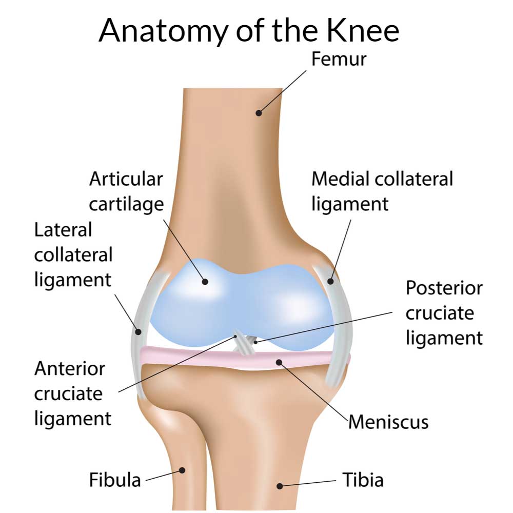
The four major ligaments that connect the upper and lower bones of the knee are:
- Anterior Cruciate Ligament (ACL)
- Posterior Cruciate Ligament (PCL)
- Medial Collateral Ligament (MCL),
- Lateral Collateral Ligament (LCL).
- Ligaments are tough, fibrous tissues. The cruciate ligaments (ACL and PCL) support the knee in the side plane and limit forward and backward motion of the knee bone. The collateral ligaments (MCL and LCL) support the knee in the frontal plane and limit side to side motion of the joint. All four of these elements of the knee anatomy work collectively to stabilize and support the knee joint. The ligaments prevent any excessive forward or backward sliding motion of the bones within the joint.
Articular cartilage is a thin layer of padding that protects the ends of each bone and cushions the bones during joint movement. The Menisci are rubbery discs that act as a shock absorbers, and facilitate the transfer forces throughout the knee joint. Menisci also ensure proper weight distribution between the bones. The medial meniscus is larger and is found on the inner side of the knee. The lateral meniscus is found on the outer side of the knee.
The sophisticated structures of the knee joint can be easily injured when unusual forces are applied during sports, accidents, overuse or even natural wear and tear. Due to the nature of the elements of the knee being connected, many injuries of the knee can affect multiple structures in the joint.
Common injuries of the knee treated by Dr. Williams include:
To obtain more information and resources on knee anatomy or common conditions of the knee, contact the office of Riley Williams, MD, orthopedic knee specialist serving Manhattan and New York City.
Locations
610 W 58th Street
New York, NY 10019
148 39th Street, 7th Floor
Brooklyn, NY 11232
Office Hours
Monday-Friday: 9:00 am – 4:30 pm
Fax: 212-774-2895