Articular Cartilage Injury Specialist

Are you experiencing knee pain and swelling with activity? If so, you may have an articular cartilage injury. Articular cartilage injuries occur when the cartilage of the knee becomes damaged and can eventually lead to osteoarthritis. Articular cartilage injury specialist, Doctor Riley J. Williams provides diagnosis as well as surgical and nonsurgical treatment options for patients in Manhattan, Brooklyn, New York City and surrounding areas who are experiencing symptoms of an articular cartilage injury. Contact Dr. Williams’ team today!
What is cartilage in the human body?
The human body has three different types of cartilage that are classified by their strength, purpose, elasticity and structure. The three cartilage types are:
- Articular Cartilage – Sometime called hyaline cartilage which covers joint surfaces at the bony ends
- Fibrocartilage – Sits between joints and acts as a cushion, examples are the meniscus, the shoulder labrum and vertebral discs
- Elastic Cartilage – Recognized for its ability to snap back into its original form, examples are the outer ear and nose.
What is articular cartilage in the knee?
Articular cartilage is a complex living tissue that lines the ends of the bones in the joint. This slick, tough tissue is often compared to the cartilage (gristle) that exists on the ends of chicken bones. The purpose of the articular cartilage is to provide a smooth surface for the bones to move against each other with a pain-free glide. The articular cartilage in the knee helps the joint withstand weight bearing through movement as it protects the bones of the joint from rubbing together. Knee articular cartilage has the ability to maintain itself. The cells in articular cartilage receive nutrients from the knee joint fluid; knee cartilage cells (chondrocytes) make the materials necessary to maintain articular cartilage structure. Cartilage does not have its own blood vessels and has a poor blood supply; the lack of blood vessels, makes the surface appear white in color. Articular cartilage is self-lubricating due to its high fluid content.
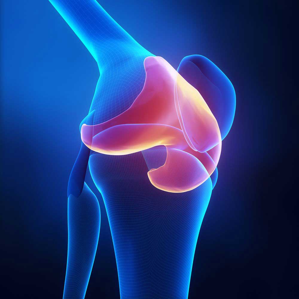
CLICK IMAGE TO ENLARGE
Articular Cartilage of the Knee
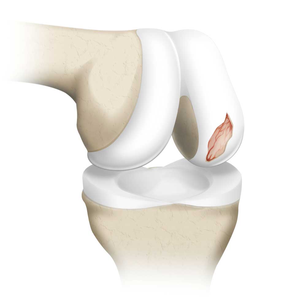
CLICK IMAGE TO ENLARGE
Articular Cartilage Injury
What is an articular cartilage injury in the knee?
An articular cartilage injury I the knee occurs when damage occurs to the tough, thin cartilage that lines the ends of the bones. Articular cartilage injuries are often caused by trauma, knee instability, or by progressive degeneration from associated with long-term wear and tear. In sports activities, a direct blow to the knee in sports like hockey, football, soccer, or Lacrosse can cause articular cartilage damage. Cartilage injuries can also occur in association with instability events (knee dislocations) such as a dislocating patella or a dislocating knee that occurs with a tear of the anterior cruciate ligament (ACL). Dr. Riley J. Williams, orthopedic knee specialist, diagnoses and treats patients in Manhattan, Brooklyn, New York City, NY and surrounding areas who have an articular cartilage injury in the knee. Dr. Williams has extensive experience with these types of knee injuries and can offer the best orthopedic treatments available.
What are the grades of an articular cartilage injury?
Orthopedic knee specialists use the Outerbridge classification system to classify the degree of articular cartilage damage. The grades are based on how much damage is present when seen during a knee arthroscopy. The grades of an articular cartilage injury are:
- Grade 0 – Normal healthy cartilage, no changes
- Grade 1 – Cartilage shows softening and inflammation
- Grade 2 – Partial thickness loss, may show surface fissures
- Grade 3 – High grade partial thickness cartilage loss – shows defect to the level of subchondral bone. Defect is in an area measuring 1.5cm or less
- Grade 4 – Full thickness cartilage loss, subchondral bone is exposed
What is a cartilage shear injury?
The type of injury on the articular surface of cartilage within the knee is influenced by the type of load sustained at the time of injury. Cartilage injuries from a hard twist with the weight of the body directly over the injury site, or a knee dislocation can cause what is called a high shear force. A sudden dislocation, or subluxation can be the cause of a cartilage shear injury. A cartilage shear injury is often seen in sports, and occurs when a piece of cartilage literally shears off the end of the end of the bone. Cartilage shear injuries are not common, but they can produce pain, swelling, locking, and a feeling that something is loose or catching within the knee joint.
What are the types of knee cartilage damage?
In general, there are two main types of knee cartilage damage involving the knee: the meniscus cartilage or the articular cartilage. There are different terms for these types of knee cartilage damage which include:
- Torn meniscus
- Frayed meniscus
- Focal articular cartilage damage
- Articular cartilage damage
- Chondral defect, lesion or damage
- OCD lesion (osteochondritis dissecans)
- Osteochondral defect, lesion or damage
- Cartilage loose body
What is a knee cartilage loose body?
After a knee injury, or prolonged wear-and-tear, the knee cartilage can become frayed or damaged. On occasion, a fragment of the articular cartilage can break away. A knee cartilage loose body is a fragment of tissue or bone that freely floats inside the knee joint space. There can be one or several loose bodies within the joint, suspended in the fluid of the knee, called the synovial fluid. Loose bodies can impact the way the knee moves and can cause symptoms such as:
- Locking of the knee joint, making it difficult to bend or straighten the knee
- Knee catching
- Crepitus – a crunching or popping sound within the knee
- Trouble walking
- Knee pain
- The feeling something is moving inside the joint
- Swelling and Inflammation
What are the symptoms of an articular cartilage injury in the knee?
Cartilage injury symptoms can be similar in nature, rather it is an articular cartilage injury or an injury of the meniscus. Symptoms may include:
- Pain
- Locking sensation in the knee
- Decreased range of motion
- Tenderness and swelling
- Feeling that the knee will “give way” or is unable to support full weight
- Inability to participate in high impact activities and sports
How are articular cartilage injuries diagnosed?
Most orthopedic knee specialists, like Dr. Riley J. Williams will use an MRI Scan to diagnose an articular cartilage injury. An MRI will show him the articular cartilage surfaces and he can determine the size, shape and progression of the injury. For some patients, especially those who cannot undergo an MRI scan, a knee arthroscopy can be an excellent diagnostic tool for helping Dr. Williams visualize the articular cartilage. He can see through an arthroscope (a small camera placed into the joint) which displays images on a television-type screen and can determine the amount and type of damage.
How is an articular cartilage injury treated?
Non-surgical treatment:
Some patients will respond well to conservative treatments for specific types of articular cartilage injuries. Non-surgical treatment may include:
- Activity modification
- Physical therapy
- NSAIDs – non-steroidal anti-inflammatory drugs
- Steroid injections
- Biological treatments which may include hyaluronic acid (gel) injections, platelet rich plasma (PRP) injections, bone marrow blood injections, or adipose (fat) based stem cell injections
Surgical treatment:
Dr. Williams has an arsenal of articular cartilage injury treatments that have been proven effective for his patients in New York. He offers the best and most up-to-date treatment options for articular cartilage injuries. These may include:
- Debridement – Smoothing the damaged cartilage and removing loose pieces
- Microfracture – Creating small holes in the base of a cartilage lesion to encourage the bone to “bleed”, heal, and create a new cartilage surface.
- Autograft Mosaicplasty – Moving healthy cartilage-bone plugs from a low demand portion of the patient’s knee to the fix a damaged area of cartilage in the same knee. This procedure is also known as autograft osteochondral transplantation (AOT).
- Osteochondral Allograft – Healthy donor cartilage-bone plugs are implanted into an area of cartilage damage to replace unhealthy cartilage. This procedure restores the natural cartilage surface and can be used for large cartilage defects.
- Matrix Associated Autologous Chondrocyte Implantation (MACI) – A two-step process that results in the creation of cartilage patch made up of the patient’s cells and a new cartilage matrix. At the first stage, a small sample of healthy cartilage is arthroscopically removed from the knee, and used to grow the new patch in an artificial environment. At the 2nd stage, the new collagen patch is placed over the damaged area, allowing new cartilage growth.
- Particulate Juvenile Articular Cartilage – This one step process utilizes small donor cartilage pieces that are placed into a cartilage defect. These cartilage “seeds” grow to form to new cartilage and fill areas of cartilage damage. This method is very effective in the femur and patella.
- Stem Cell Injections: Stem cells obtained from the bone marrow of the hip (iliac crest) or adipose (fat) can be used in association with any of the described methods of cartilage repair above to aid in the healing and successful reconstruction of treated articular cartilage defects.
For more information on articular cartilage injuries, knee cartilage repair, or on the many different options available for your knee cartilage injury, please contact the office of Riley Williams, MD, knee specialist serving Manhattan, Brooklyn, New York City, NY and surrounding areas.
Locations
610 W 58th Street
New York, NY 10019
148 39th Street, 7th Floor
Brooklyn, NY 11232
Office Hours
Monday-Friday: 9:00 am – 4:30 pm
Fax: 212-774-2895