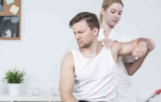
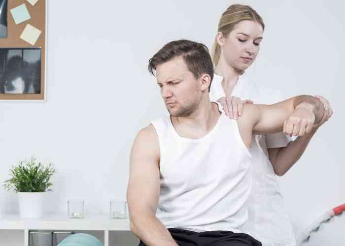
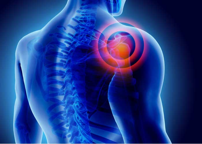
Shoulder Impingement Specialist

Do you participate in activities at work or in sports that involve throwing overhead? If so, you may be at risk of developing shoulder impingement. One of the most common causes of shoulder pain is shoulder impingement (sometimes call subacromial impingement.) Shoulder impingement specialists, Doctor Riley J. Williams provides diagnosis as well as surgical and nonsurgical treatment options for patients in Manhattan, Brooklyn, New York City and surrounding areas who are experiencing shoulder pain and shoulder impingement. Contact Dr. Williams’ team today!
What is shoulder impingement?
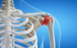
Shoulder impingement, also known as swimmer’s shoulder or impingement syndrome, is a common cause of shoulder pain. Shoulder impingement is caused by the compression of the rotator cuff tendons between the acromion (highest portion of the shoulder blade) and the humerus (upper arm). The result of this process is inflammation within the subacromial space, and subsequent thickening of the tendons and bursa (fluid-filled sacs). With the thickening of the rotator cuff tendons, a decrease in the amount of space available for tendon movement occurs, this lack of functional space results in the pinching of the rotator cuff tendons and bursae between the bones. Dr. Riley J. Williams, orthopedic shoulder specialist serving patients in Manhattan, Brooklyn, New York City, NY and surrounding areas, has the knowledge and understanding, as well as substantial experience in treating patients who have experienced shoulder impingement.

What is subacromial impingement?
Subacromial impingement is characterized by the irritation and inflammation of the tissue the sit just below the acromion process (top of the shoulder). The rotator cuff tendon and bursa exist in this space. When affected individuals raise their arm overhead, these structures are compressed between the upper arm and top of the shoulder blade. The pain associated with subacromial impingement typically occurs with elevation of the arm above the head due to the narrowing of the subacromial space.
What causes shoulder impingement?
Shoulder impingement is commonly the result of repetitive overhead actions or chronic shoulder use. Inflammation of the bursa (bursitis) can occur from a number of injuries. The thickening of the tendons from continuous irritation can generate mechanical symptoms such as rubbing, grinding, or pinching. This is common among young athletes who perform repetitive overhead actions in sports like volleyball, baseball, tennis, and swimming. Individuals working in laborious type jobs (construction, building, painting), or someone who repetitively lifts overhead are also prone to shoulder impingement.
What are the symptoms of shoulder impingement?
The most common symptoms of shoulder impingement are:
- Sharp pain with activities involving reaching overhead or behind the back
- Radiating pain from the shoulder into the arm
- Shoulder weakness
- Worsening pain at night, particularly while sleeping on the affected side
- Aching pain noted at the upper arm
How is shoulder impingement diagnosed?
Prompt medical attention is recommended if shoulder impingement is suspected. Because the tissue will continue to thicken with repeated shoulder use, shoulder pain can worsen and damage to the rotator cuff can occur. If treated early, patients can minimize swelling and continued discomfort using conservative methods. Dr. Williams can diagnose shoulder impingement with a comprehensive medical history and physical examination. Diagnostic testing, including x-rays and magnetic resonance imaging (MRI), may be requested to identify any damage to any other structures within the shoulder.
What is the treatment for shoulder impingement?
Non-surgical treatment:
Patients who have been diagnosed with shoulder impingement are typically able to recover with conservative therapy. Activity modification, non-steroidal anti-inflammatory medications, and shoulder exercises can help patients reduce shoulder pain and inflammation associated with shoulder impingement. If the pain is not relieved by oral medications, a corticosteroid or platelet rich plasma (PRP) injection directly into the subacromial space can also be employed. When the pain and swelling have decreased, Dr. Williams will typically recommend a physical rehabilitation program aimed at strengthening the tendons and muscles of the rotator cuff and shoulder blade.
Surgical treatment:
If conservative methods are unsuccessful, Dr. Williams may recommend shoulder arthroscopy; this procedure is a minimally invasive surgery that uses a small camera to view the tendons and muscles of the shoulder. During this procedure, any irregularities, such as bone spurs or thickened bursa, are removed. Noted tears are repaired using specialized surgical instruments. A treatment called subacromial decompression will eliminate compression of the tendons between the humerus and the acromion. Dr. Williams does this procedure utilizing small incisions, thereby allowing patients to return to their normal activities in a shorter period of time.
For more information on shoulder impingement, subacromial impingement, or the excellent treatment options available, please contact the office of Dr. Riley J. Williams, orthopedic shoulder specialist serving Manhattan, Brooklyn, New York City, NY and surrounding areas.
Subacromial Decompression Doctor
Do you participate in sports or in work that involves continuous overhead motions, or throwing overhead? If so, you may be at risk of developing subacromial impingement. Subacromial decompression surgeon, Doctor Riley J. Williams provides diagnosis as well as surgical and nonsurgical treatment options for patients in Manhattan, Brooklyn, New York City and surrounding areas who are experiencing subacromial impingement and related shoulder pain. Contact Dr. Williams’ team today!
What is shoulder impingement?
Shoulder impingement is one of the most common causes of shoulder pain. Shoulder impingement caused by compression of the rotator cuff between the acromion (highest portion of the shoulder blade) and humerus (upper arm) during overhead activities. This compression results in propagation of inflammation within the subacromial space. This inflammation causes thickening of the rotator cuff tendons and bursa. The thickening of the rotator cuff tendons, and the thickening of the bursa decreases the space available for tendon movement, thereby causing compression of the tendons and bursae between the bones.

What is a subacromial decompression?
Within the shoulder, there are muscles that allow the shoulder to rotate, tendons that stabilize the shoulder with movement, and fluid-filled sacs (bursa) that help cushion shoulder movement. The muscles of the shoulder travel under the acromion (subacromial), and continue to the humerus (upper arm bone). In patients who perform repetitive overhead activities, the rotator cuff tendons consistently rub against the acromion; this rubbing causes inflammation of the tendons and bursa and decreases the space available for normal shoulder movement. Patients who experience shoulder pain and weakness due to impingement and bursitis may be candidates for a surgical procedure called a subacromial decompression. The goal of this arthroscopic procedure is to create space below the acromion to facilitate normal shoulder motion. Dr. Riley J. Williams, orthopedic shoulder surgeon, treats patients in Manhattan, Brooklyn, New York City, NY and surrounding areas who have suffered from shoulder impingement and are in need of subacromial decompression.
How is subacromial decompression performed?
Dr. Williams uses an arthroscopic approach to this procedure. Arthroscopy utilizes small incisions in the shoulder that allow access to the affected areas. During an arthroscopic subacromial decompression, a small camera (arthroscope) is inserted into the shoulder to visualize the subacromial space. Any irregularities such as tears, bone spurs, inflamed bursa, or irregular tissues are removed using specialized surgical instruments. The removal of pathologic tissue allows the muscles and tendons to heal and reduces the irritation with shoulder movement.
What other procedures can be with a subacromial decompression?
Dr. Williams prefers the arthroscopic method for subacromial decompression because it is minimally invasive. Due to the size of the camera and specialized surgical instruments, the incisions are small and allow for a faster recovery time for the patient. Arthroscopic subacromial decompression also reduces the risk of infection, blood loss, as well as decreased pain and inflammation. Oftentimes the acromioclavicular (AC) joint can be visualized during a subacromial decompression. In patients with arthritis of the AC joint, the distal clavicle can be trimmed to remove the arthritic portions of the AC joint and create space between the collarbone and shoulder blade. This arthroscopic procedure can provide significant pain relief in this area.
What is the recovery period like after subacromial decompression with or without AC resection?
Although the recovery period is variable, most patients can expect to return to daily routine within 2-3 weeks. Sporting activities can resume within 2-4 months following a successful subacromial decompression. In general, patients in New York can expect the following:
- A sling is applied to immobilize the shoulder joint immediately following surgery.
- Active movement of the shoulder and arm begins immediately after surgery.
- Dr. Williams will provide specific shoulder exercises to achieve full range of motion within 2-4 weeks following surgery.
- Formal physical therapy is usually necessary for 2-3 months
For more information on subacromial decompression, or to find a treatment for shoulder impingement, please contact the office of Riley J. Williams, MD, an orthopedic shoulder surgeon serving Manhattan, Brooklyn, New York City, NY and surrounding areas.
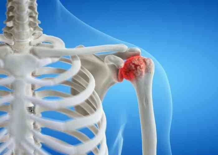
Shoulder Arthritis Specialist
Are you experiencing mild to severe shoulder pain that feels worse after activity or exercise? If so, you may have shoulder arthritis. Arthritis of the shoulder is typically found in middle-aged adults and the older population and is caused by the natural aging process. Shoulder arthritis specialist, Doctor Riley J. Williams provides diagnosis as well as surgical and nonsurgical treatment options for patients in Manhattan, Brooklyn, New York City and surrounding areas who are experiencing the symptoms of shoudler arthritis. Contact Dr. Williams’ team today!
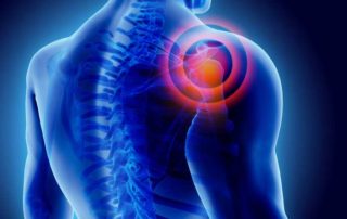
What is shoulder arthritis?
Although the shoulder is comprised of four joints, the main joint is the glenohumeral joint . The glenohumeral joint describes the articulation of the humeral head (ball) and glenoid (socket). The ends of these bones are lined with protective connective tissue (cartilage), which allow the joint to move smoothly. The other joints of the shoulder include the acromioclavicular (AC) joint, the the sternoclavicular joint (SC) and the scapulothoracic joint. The SC joint is where the clavicle (collarbone) and sternum meet, and the AC joint is where the clavicle and scapula (shoulder blade) meet on top of the shoulder. When cartilage is damaged, arthritis results, which leads to bone-on-bone rubbing. This painful friction can limit mobility, range of motion, and cause pain. Shoulder arthritis can occur in any of these joints; glenohumeral joint arthritis is a serious clinical issue. If the arthritis causes extensive damage, total shoulder replacement, or total shoulder arthroplasty, may be recommended. Dr. Riley J. Williams, orthopedic shoulder specialist serving Manhattan, Brooklyn, New York City, NY and surrounding areas has extensive experience in treating shoulder arthritis and shoulder related injuries.

What are the symptoms of shoulder arthritis?
Arthritis in the shoulder typically occurs over time and slowly wears down the cartilage within the joint(s). Cartilage softens, cracks, flakes, and erodes over time. Cartilage loss results in a bone-on-bone situation.
This can cause:
- Pain
- Irritation
- Swelling
- Stiffness & motion loss
- Redness of the skin at the site of irritation
- Crepitus (cracking noises)
How is shoulder arthritis diagnosed?
The most common form of arthritis of the shoulder is osteoarthritis (wear and tear arthritis). Rheumatoid arthritis is an autoimmune disease that can cause an inflammatory response in the shoulder. Avascular necrosis (bone death) is a condition that is caused by a disruption of blood supply, leading to arthritis. Shoulder dislocations from trauma (fall, car accident or sports accident) can also lead to an arthritic condition. Chronic tearing of the rotator cuff can lead to shoulder arthritis (rotator cuff arthropathy). Dr. Williams will discuss your medical history and shoulder injury and perform a physical exam. The severity of the shoulder arthritis can be determined with imaging tests such as x-rays, CT scans or MRIs.
How is shoulder arthritis treated?
Non-surgical treatment:
Dr. Williams will help determine what conservative treatment methods will be beneficial to your condition. Daily range of motion exercises and stretching can help keep the shoulder joint mobile. An individual may need to modify his or her lifestyle, and decrease or cease activities that aggravate shoulder pain. Over the counter pain medication, such as ibuprofen or naproxen, can help treat arthritis flare-ups as well as help decrease inflammation. Icing the injured shoulder can also help provide relief from pain and inflammation. Shoulder injections (corticosteroids, platelet rich plasma) can also help manage arthritic shoulder pain.
Surgical treatment:
If arthritis related shoulder pain and loss of mobility progress and non-surgical methods are ineffective, surgery may be recommended. There are several different types of surgery that can be performed depending on the severity of the arthritis. Arthroscopic shoulder debridement is a type of procedure that essentially cleans out the joint and provides temporizing provide relief. Occasionally, bone spurs can form in the shoulder and surgery to shave down the excess bone can help improve pain and restore mobility. AC joint arthritis can be treated arthroscopically by partially resecting the most distal portion of the clavicle to create room between the bones. In general, arthroscopic management strategies can delay the need for total shoulder replacement. In the most severe cases an individuals suffering glenohumeral joint arthritis may need total shoulder replacement surgery . During total shoulder replacement, the ball and socket portions of the shoulder joint are resurfaced with metal and plastic prostheses. As a leading orthopedic shoulder specialist in New York, Dr. Williams will discuss the severity of the injury and which surgical treatment options are best for improving shoulder pain and mobility.
For more information on shoulder arthritis and the treatment options available, please contact the office of Riley J. Williams, MD, orthopedic shoulder specialist serving Manhattan, Brooklyn, New York City, NY and surrounding areas.
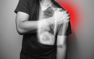
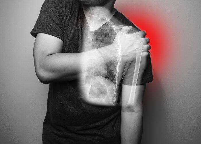
Broken Shoulder Specialist
Do you play contact sports? If so, you may be at risk of sustaining a shoulder fracture. A shoulder fracture can be caused by a sudden force, impact or trauma to the joint during sports activities. A broken shoulder can also occur from a hard fall or accidnet. Broken shoulder specialists Doctor Riley J. Williams provides diagnosis as well as surgical and nonsurgical treatment options for patients in Manhattan, Brooklyn, New York City and surrounding areas who have sustained shoulder fracture. Contact Dr. Williams’ team today!
What is a shoulder fracture?
The shoulder is a ball and socket joint and consists of three bones: the humerus (upper arm bone) clavicle (collarbone) and scapula (shoulder blade). A fracture is a break in any one of the shoulder bones. Fractures occur in association with traumatic events like falls, car accidents or sports related impacts. There are two types of shoulder fractures: displaced and non-displaced. Displaced fractures may require surgery to reconstruct the affected bone and enable the return of normal function. The majority of shoulder fractures are non-displaced, which means the bones fragments are near their proper position; surgery is usually not necessary. Dr. Riley J. Williams, orthopedic shoulder specialist serving Manhattan, Brooklyn, New York City, NY and surrounding areas has extensive experience in treating shoulder fractures and shoulder related injuries.

What are the symptoms of a shoulder fracture?
Individuals in the New York area who have experienced trauma from a fall, a motor vehicle accident, a direct blow to the shoulder from sports may report the following symptoms in association with a shoulder fracture:
- Pain
- Swelling
- Bruising
- Limited mobility (if any)
- Grinding or clicking feeling
- Obvious deformity of the shoulder
How is a shoulder fracture diagnosed?
There are a few different types of breaks an individual may sustain in the shoulder. One type of fracture is a proximal humerus fracture. This type of break affects the ball portion of arm bone (humeral head). Elderly individuals are at risk for this type of break due to osteoporosis, which weakens bones. A fall on an outstretched hand can be cause for a proximal humerus fracture. A proximal humerus fracture can be simple or complex (comminuted). An x-ray can determine the type of proximal humerus fracture an individual has sustained. A scapula fracture is a break to the shoulder blade; this is an uncommon break due to its location at the upper rear thorax. A clavicle fracture is a common type of break of the collarbone. The clavicle connects the shoulder blade to the ribcage; the collarbone is at risk for fracture during a fall. Clavicle fractures occurs during many sporting activities such as football, soccer, cycling, downhill skiing, rugby and lacrosse. Dr. Williams will ask questions about the injury and perform a physical examination of the shoulder. An x-ray or musculoskeletal ultrasound will determine if there is a broken bone. Other imaging tests like a CT Scan or MRI may be necessary if other injuries were sustained with the break.
How is a shoulder fracture treated?
Non-surgical treatment:
Conservative treatment is typically reserved for non-displaced fractures. This means the bones have not shifted within the shoulder and are still aligned in an acceptable position. Bracing, sling wear, and immobilization can be recommended in cases of nondisplaced shoulder fractures. Dr. Williams may prescribe physical therapy during the healing process so that an individual can regain range of motion and shoulder function. Icing the injury and taking over-the-counter medication, such as ibuprofen or naproxen, can help with swelling and pain.
Surgical treatment:
Traumatic shoulder fractures and displaced fractures can require surgery. The goal of surgery is the proper positioning of a displaced fracture to an anatomic position. The surgical treatment necessary depends on the type of shoulder injury and type of fracture that was sustained. Fracture repair (open reduction and internal fixation) involves the use of plates and screws to facilitate proper bony healing; in the most severe cases of proximal humerus fractures, partial or total shoulder replacement surgery may be necessary. Dr. Williams will discuss the severity of the injury sustained and what surgical procedure are best for the patient to resolve the issue. Surgical treatment will also require physical therapy sessions after an individual has recovered from the procedure and the bone has begun to heal.
For more information on a shoulder fracture, including a proximal humerus fracture and a scapula fracture and the advanced treatment options available, please contact the office of Riley J. Williams, MD, orthopedic shoulder specialist serving Manhattan, Brooklyn, New York City, NY and surrounding areas.
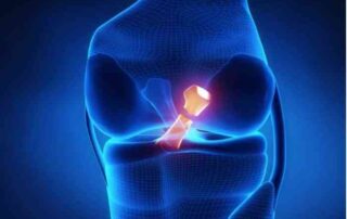
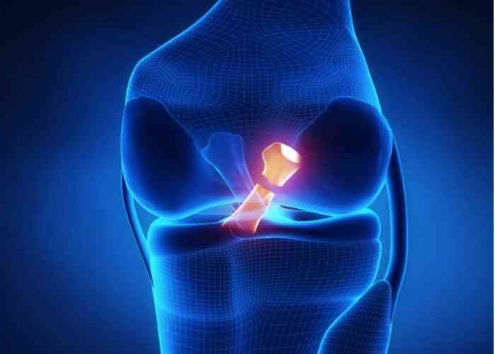
ACL Revision/Reconstruction Doctor
Are you an athlete who participates in sports that involve jumping or quick stopping? If so, you may had had an ACL reconstruction for a torn anterior cruciate ligament. Occasionally, ACL reconstruction fails and a new ACL revision or reconstruction surgery is needed to restore the function of the knee. ACL revision and reconstruction surgeon, Doctor Riley J. Williams provides diagnosis as well as the most up-to-date surgical options available for patients in Manhattan, Brooklyn, New York City and surrounding areas who have sustained an ACL re-injury or who have a failed ACL surgery. Contact Dr. Williams’ team today!
What is a revision ACL reconstruction?
The anterior cruciate ligament (ACL) is a commonly torn ligament in the knee. This ligament connects the femur (thighbone) to the tibia (shinbone) and helps to stabilize the knee. ACL tears and injuries are most often experienced by athletes participating in sports that involve jumping, twisting, and changing direction (soccer, basketball, lacrosse, downhill skiing). Patients who experienced a severe ACL injury will typically undergo ACL reconstruction surgery; ACL reconstruction repairs the damage and restores normal function to the knee joint. ACL reconstruction surgery has a very high rate of success. However, ACL tears can reoccur. Athletes who return to pivoting sports are at risk for recurrent ACL tear even following a successful rehabilitation. In these cases, revision ACL reconstruction is required to restore the knee and to repair the damage and restore functional knee stability. Dr. Riley Williams, a complex knee surgeon, successfully performs revision ACL reconstruction for patients in Manhattan, Brooklyn, New York City, NY and surrounding areas who have experienced failure after a recent ACL reconstruction.

What causes an ACL reconstruction to fail?
The goal of ACL reconstruction is to restore knee stability, and to restore normal knee function. Failure of an ACL reconstruction occurs when the patient experiences a tear of the reconstructed ligament or if the knee is unable to function properly after the index procedure. ACL reconstruction failure may result in knee pain, loss of range of motion, or recurrent knee instability. ACL reconstructions may fail for a variety of reasons. The procedure may have been performed before normal range of motion is restored to the injured knee; these circumstances may result in knee loss of motion following surgery. MRI of such knees often demonstrate the presence of “cyclops lesion” or scar tissue in the front of the knee. Fortunately, removal of this scar tissue can alleviate this problem and allow for a full recovery. Other causes of ACL surgery failure include failure to adequately follow the post-operative rehabilitation plan, technical issue with ACL graft placement, and new trauma to the ACL-reconstructed knee.
Who should have a revision ACL reconstruction?
Most active patients who suffer a recurrent ACL tear will likely need a revision reconstruction. Dr. Williams will discuss the pros and cons of such a procedure and how best to optimize the clinical results following this surgery. Dr. Williams believe that it is crucial to understand the causes of the failed ACL procedure in order to avoid similar issues following the revision reconstruction. It is important to follow the post-operative plan delineated to the patient by Dr. Williams. Following surgery, it is normal to experience some swelling, pain, and decreased range of motion while in the acute healing phase. Over time, patients are advised to begin strength exercises and more stretching. This process helps to gradually regain strength and flexibility in the knee and may take a few months to complete. After about six months, the knee should be fully functional and stable.
Revision ACL reconstruction surgery is an excellent choice for patients who have undergone primary ACL reconstruction surgery and experienced another injury. If the patient returns to sports or weight-bearing activities too quickly following surgery, they run the risk of tearing the new tendon and causing further damage to the knee joint. In general, if patients expect to return to pivoting sports, revision ACL reconstruction will be necessary to achieve this desired goal.
How is revision ACL reconstruction performed?
Before revision ACL reconstruction surgery is performed, patients meet with Dr. Williams to discuss the cause of the ACL graft failure. A full understanding of the graft failure is needed so that the risk of repeat failure the revision surgery is minimized. Revision ACL reconstruction is a similar procedure to primary ACL reconstruction. Dr. Williams prefers the use of the patient’s tissue (autograft) for revision ACL reconstruction as these tissues are very strong and heal quickly and predictably.
During the first reconstruction, tunnels were created in the tibia and femur bones to facilitate ACL graft placement. If these tunnels have been damaged or expanded, bone graft may be used to repair the bone defect so the new tendon graft can be secured in place. In some cases, the original ACL graft may have been placed in the wrong footprint; if this is the case, the bony portion of the original graft may be left in place. Dr. Williams will do that which is necessary to place the revised ACL ligament graft into the proper location within the knee joint.
Sometimes, revision ACL reconstruction requires two separate surgeries. The first operation is performed to restore the bony integrity of the knee joint. Bone plugs and bone graft are used to restore the bony anatomy of the tibia and femur. Reconstitution of the tibia and femur allows for the placement of the revision ACL graft in the correct anatomic location. After about 3-6 months, Dr. Williams will check the graft procedure using a CT or MRI scan. Once the bony repair is physiologically complete, Dr. Williams will perform a second surgery to place the new ACL graft. During this operation, Dr. Williams creates new tunnels in the tibia and femur to secure the graft. He takes care to ensure that the new grafts are in the correct anatomical position and will allow the optimal range of motion and stability.
Revision ACL graft sources include use of a hamstring tendon autograft, quadriceps tendon autograft, and contralateral patellar tendon graft. The patient’s own tissue is Dr. Williams’ preferred revision graft of choice. In some cases, donor tissue (allograft) may be indicated. Dr. Williams will discuss all these options with the patient prior to performing the revision ACL procedure.
In addition, adjunctive procedures such as a lateral extra-articular tenodesis, iliotibial band tenodesis, or anterior lateral ligament (ALL) may be recommended by Dr. Williams to increase the likelihood of clinical success of revision ACL reconstruction.
How long is the recovery after revision ACL reconstruction?
In general, the recovery is the same as primary ACL reconstruction: 6 months. Bracing is used for 6 weeks after surgery. Physical therapy starts one week following surgery and continues for approximately 3-4 months following surgery. Strength and conditioning is very important near the end of the initial six-month postoperative interval. Patients should expect a measure approach to a full return to play. A full functional recovery must be established prior to a return to sports and full activity. Dr. Williams recommends the use of objective strength measures, movement analysis and an assessment psychological readiness prior to full clearance following revision ACL reconstruction. It is important not to rush the recovery and rehabilitation process after revision ACL reconstruction. Recovery will often take longer the first reconstruction. The goal is to optimize stability and movement without damaging the graft or knee joint. In general, patients can expect to return to their athletic activities approximately 9 months after surgery.
For more information about revision ACL reconstruction or the causes of ACL reconstruction failure and the best treatment options available, please contact the office of Riley J. Williams, MD, orthopedic knee surgeon serving Manhattan, Brooklyn, New York City, NY and surrounding areas.