Knee Osteotomy Surgeon

Are you experiencing pain and stiffness in your knee? Have you been previously diagnosed with malalignment syndrome? If so, you may have arthritis in your knee. A knee osteotomy is generally reserved for patients who have damage on just one side of the joint. Knee osteotomy surgeon, Doctor Riley J. Williams provides diagnosis as well as surgical and nonsurgical treatment options for patients in Manhattan, Brooklyn, New York City and surrounding areas who require a knee osteotomy. Contact Dr. Williams’ team today!
What is a knee osteotomy?
A knee osteotomy is a surgical procedure that is performed to correct malalignment of the bones of the knee. Dr. Riley J. Williams, orthopedic knee surgeon serving patients in Manhattan, Brooklyn, New York City, NY and surrounding areas is extremely skilled at performing different types of knee osteotomies.
During an osteotomy procedure, Dr. Williams partially cuts a bone that is in a mechanically poor position. This cut allows him to realign or move the bone into a better mechanical position. The realigned bone is often stabilized with strong metal plates and screws.
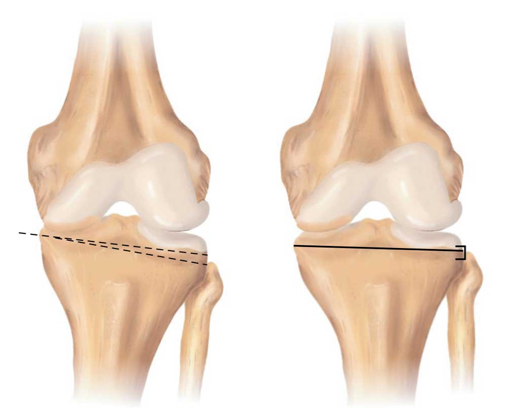
CLICK IMAGE TO ENLARGE
Knee Osteotomy
What is knee malalignment syndrome?
The knee is comprised of three bones: the femur (thighbone), tibia (shinbone) and patella (kneecap). It also contains cartilage, tendons and ligaments. There are two joints within the knee: the tibia-femur joint and the patella-femur joint. Knee malalignment syndrome occurs when there is poor positioning between either of these two knee joints. In knee malalignment of the patella-femur joint, the kneecap feels either unstable, or feels too tight and painful. In knee malalignment of the tibia-femur joint, abnormal knee angulation can create undue pressure on either the inner (medial) or outer (lateral) compartment of the knee. Knee malalignment can put individuals in New York at higher risk for knee problems including knee dislocation and osteoarthritis.
Tibia-femur malalignment can give the appearance of genu valgum (knock knee) or genu varus (bow-legged). Genu valgum displays when an individual is standing straight up, their knees are together, and their feet and ankles appear spread apart. Genu varus is shown when an individual is standing straight up, their knees are apart (lower legs curve outward from the knee), and their feet appear to touch. Genu valgum and genu varum can occur as a result of birth related issues, follow a traumatic accident, or can be associated with the aging process. Dr. Williams has extensive experience in diagnosing and treating knee malalignment.

What are the symptoms of a knee malalignment syndrome?
Knee pain may worsen with normal daily activities. Sports like running, or jogging may be very difficult. Simple tasks like squatting, sitting for long periods of time, or going up and down stairs. Symptoms may include:
- Knee stiffness & swelling
- Grinding or clicking sounds when the knee is extended
- Knee giving out unexpectedly
- Pain under the kneecap, inner knee, or outer knee
- Knee locking or catching
- Excessive angulation of the knee
What is a Tibial Tubercle Osteotomy?
The patellar tendon connects the kneecap (patella) to the lower leg (tibia); the attachment site of the patellar tendon on the tibia is called the tibial tubercle. Malalignment or poor positioning of the tibial tubercle can cause patellar instability (slipping or dislocation) or patellar overload syndrome (high joint pressure causing pain). A tibial tubercle osteotomy involves moving the tibial tubercle to a more normal position on the tibia. Typically, a small incision is made just over the anterior tibia, just below the knee joint. The tibial tubercle is exposed and moved along with a small segment of bone. The moved segment of bone is held in place using two metal screws. In cases of patellar instability, the tibial tubercle is moved medially (toward the inner knee); this corrects the abnormal angle between the tibia and femur that leads to dislocations (Q-angle). In cases of patellar overload, the tibial tubercle is moved anteriorly (forward); this elevates the patellar tendon and the patella so as to decrease the joint forces on the kneecap. This reduction in pressure results in pain relief and better tracking of the patellar in the trochlear groove of the femur.
Tibial tubercle osteotomy is typically done on an outpatient basis. The procedure takes approximately 30 minutes and may be combined with other procedures (cartilage repair, medial patellofemoral ligament repair). Patients usually wear a brace for 4-6 weeks after surgery. Crutches are recommended for 1-2 week after surgery. Most patients are able to resume normal walking without the brace after six weeks. Full return to athletics and sporting activities typically occurs between 3-6 months.
What is a High Tibial Osteotomy (HTO)?
A high tibial osteotomy (HTO) is performed to treat malalignment of the tibio-femoral joint. Disorders of the tibio-femoral joint include genu varum (bow leggedness) or genu valgum (knock knees). These condition results in overload of either the inner (medial) or outer (lateral) knee joint. HTO is usually used to treat excessive bow leggedness and disorders of the inner or medial knee joint. An HTO involves making a partial cut in the proximal tibia bone just below the knee joint. This partial cut allows the surgeon to move the lower leg into a straighter more mechanically sound position. Typically, an incision is made on the inner side of the knee, below the knee joint. The bone cut is made, and the leg is moved into its new position. The newly aligned bone is held in its new position using a strong metal plate and screws. The procedure takes approximately 45 minutes and may be combined with other procedures (cartilage repair, knee ligament reconstruction). Straightening the lower extremity using an HTO results in a reduction in pressure on the affected knee compartment, patients thus experience pain relief and better overall knee function.
High tibial osteotomy is typically done on an outpatient basis; some patients may wish to spend a night in the hospital. Patients usually wear a brace for 4-6 weeks after surgery. Crutches are recommended for approximately 2-3 weeks after surgery. Most patients are able to resume normal walking without the brace after six weeks. Full return to athletics and sporting activities typically occurs between 4-6 months.
What is a Distal Femoral Osteotomy?
A distal femoral osteotomy (DFO) is performed to treat valgus (knock knee) malalignment of the tibio-femoral joint. This condition results in an overload of the outer (lateral) knee joint. A DFO involves making a partial cut in the lower thigh bone (femur) just above the knee joint. This partial cut allows the surgeon to move the lower leg into a straighter more mechanically sound position. Typically, an incision is made on the outer side of the knee, above the knee joint. The bone cut is made, and the leg is moved into its new position. The newly aligned bone is held in its new position using a strong metal plate and screws. The procedure takes approximately 45 minutes and may be combined with other procedures (cartilage repair, knee ligament reconstruction). Straightening the lower extremity using a DFO results in a reduction in pressure on the lateral knee compartment, patients thus experience pain relief and better overall knee function.
Distal femoral osteotomy can be done on an outpatient basis; some patients may wish to spend a night in the hospital. Patients usually wear a brace for 4-6 weeks after surgery. Crutches are recommended for approximately 2-3 weeks after surgery. Most patients are able to resume normal walking without the brace after six weeks. Full return to athletics and sporting activities typically occurs between 4-6 months.
For more information on knee osteotomies including tibial tubercle osteotomy (TTO), high tibial osteotomy (HTO), and distal femoral osteotomy (DFO), knee malalignment, and the treatment options available, please contact the office of Riley J. Williams, MD, orthopedic knee specialist serving Manhattan, Brooklyn, New York City, NY and surrounding areas.
Locations
610 W 58th Street
New York, NY 10019
148 39th Street, 7th Floor
Brooklyn, NY 11232
Office Hours
Monday-Friday: 9:00 am – 4:30 pm
Fax: 212-774-2895